

Breast cancer remains one of the most commonly diagnosed cancers among women worldwide. Early detection plays a crucial role in improving survival rates and expanding treatment options. Medical imaging technologies, such as breast ultrasound, help doctors evaluate abnormalities in the breast and determine whether further testing is needed.
Understanding how ultrasound helps detect potentially malignant breast lesions can empower patients to seek medical attention early and stay proactive about their breast health.
What Is a Breast Ultrasound?
A breast ultrasound is a diagnostic imaging procedure that uses high-frequency sound waves to produce images of the internal structures of the breast. Unlike some other imaging tests, ultrasound does not use radiation, making it a safe and commonly used method for evaluating breast abnormalities.
Doctors often recommend breast ultrasound for women under the age of 40 who are beginning their breast cancer screening. It may also be advised when a lump is detected during a physical exam or when a suspicious area appears on another imaging test. In addition, breast ultrasound is particularly helpful for women with dense breast tissue, where certain abnormalities may be more difficult to detect through mammography alone.
While ultrasound is not typically used as a primary screening tool for breast cancer, it serves as an important complementary imaging method that provides additional detail.
Ultrasound Results That May Suggest Malignancy
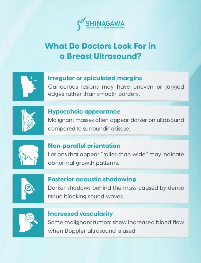
When radiologists examine breast ultrasound images, they look for specific characteristics that may indicate whether a mass is benign or potentially malignant.
Some ultrasound features that may raise suspicion for malignancy include:
- Irregular or spiculated margins – cancerous lesions may have uneven or jagged edges rather than smooth borders.
- Hypoechoic appearance – malignant masses often appear darker on ultrasound compared to surrounding tissue.
- Non-parallel orientation – lesions that appear “taller-than-wide” may indicate abnormal growth patterns.
- Posterior acoustic shadowing – darker shadows behind the mass caused by dense tissue blocking sound waves.
- Increased vascularity – some malignant tumors show increased blood flow when Doppler ultrasound is used.
It is important to note that these features alone do not confirm cancer. Instead, they help radiologists determine whether further investigation is necessary.
Benign vs. Malignant Findings: Key Differences
Many breast lumps detected on imaging turn out to be benign conditions, such as cysts or fibroadenomas. Ultrasound helps differentiate between these non-cancerous findings and lesions that may require closer evaluation.
Benign breast masses often appear with the following characteristics:
- Smooth and well-defined borders
- Oval or round shape
- Parallel orientation (wider than tall)
- Uniform internal structure
In contrast, potentially malignant masses may appear irregular, have indistinct borders, and display uneven internal patterns. Radiologists analyze the combination of these imaging features to classify lesions and determine the next steps in diagnosis.
Why Ultrasound Alone Is Not Enough
Although breast ultrasound is a valuable diagnostic tool, it cannot definitively diagnose breast cancer on its own. Imaging results must be interpreted alongside other clinical information, including physical examination findings and additional imaging tests.
In some cases, doctors may recommend further evaluation through mammography, magnetic resonance imaging (MRI), or a biopsy to confirm whether a suspicious lesion is cancerous.
Because of these limitations, ultrasound should be viewed as part of a comprehensive breast evaluation rather than a standalone diagnostic method.
People Also Ask: Conversational FAQs
Malignant breast cancer often appears as a hypoechoic, irregular solid mass with spiculated margins, taller-than-wide orientation, posterior acoustic shadowing, and increased vascularity.
A simple cyst looks smooth, fluid-filled, and dark on ultrasound, while a malignant tumor is solid, irregular, and requires biopsy for confirmation.
Ultrasound is not a replacement but a supplement to mammography. It is especially useful in women with dense breast tissue and for guiding biopsies.
The Importance of Early Detection
Detecting breast cancer at an early stage significantly improves treatment outcomes and survival rates. Smaller tumors that are identified early are often easier to treat and may require less aggressive therapies.
Patients should remain aware of possible breast changes, including:
- A new lump or thickening in the breast
- Changes in breast shape or size
- Skin dimpling or redness
- Nipple discharge or inversion
Get your Health Checkup at Shinagawa Diagnostic
In the Philippines, breast cancer ranks among the leading cancers affecting women, making it especially important for those at risk to undergo regular screening. Supporting women in practicing early detection and prevention, Shinagawa Diagnostic offers breast ultrasound both as a standalone service and as part of its specialized health package.
Through the Women’s Prime Executive Checkup, patients can undergo breast ultrasound along with other essential tests and services designed to provide a more comprehensive view of their overall health. This approach helps ensure that potential concerns are identified early while giving women the information they need to take proactive steps toward better health.