
With more than 30 years of experience, Shinagawa Lasik & Aesthetics has risen through the ranks to become a world-renowned leader in LASIK and Aesthetics with 47 branches all over Japan and two (2) in the Philippines.
Now, Shinagawa extends its expertise beyond vision care through through Japanese-standard healthcare at Shinagawa Diagnostic & Preventive Care. This clinic brings Japan’s trusted approach to health—focused on early detection and prevention—to Filipinos seeking complete wellness.
Ophthalmology and Eye Care Services
Vision Check (Eye Test)

Tonometry

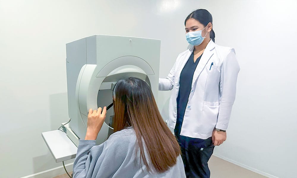
Optical Coherence Tomography (OCT)


Fundus
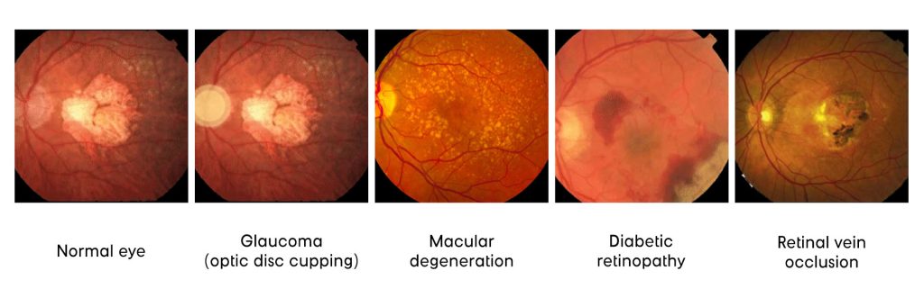
Eye Diseases Detectable Through a Fundus Examination
- Glaucoma (Optic Nerve Cupping) – A condition that damages the optic nerve, the part of the eye that carries visual information to the brain. An early sign is optic nerve cupping, where the center of the optic nerve enlarges due to increased pressure inside the eye.
- Common causes/risk factors: Increased eye pressure, aging, family history of glaucoma, diabetes, and long-term steroid use.
- Diabetic Retinopathy (Retinal Bleeding) – A complication of conditions such as diabetes that affects the blood vessels in the retina, the light-sensitive tissue at the back of the eye. High blood sugar levels can damage these vessels, causing them to leak or bleed, which may lead to blurred vision or vision loss over time.
- Common causes/risk factors: Poorly controlled diabetes, high blood pressure, high cholesterol, and long duration of diabetes.
- Macular Degeneration – A condition that affects the macula, the central part of the retina responsible for sharp and detailed vision. As the macula deteriorates, it can cause difficulty seeing fine details, reading, or recognizing faces.
- Common causes/risk factors: Aging, smoking, family history, high blood pressure, and prolonged exposure to UV light.
- Retinal Vascular Arteriosclerosis / Occlusion (Retinal Bleeding) – This condition occurs when the blood vessels in the retina become narrowed, hardened, or blocked, reducing blood flow to the eye. When the vessels are damaged or obstructed, bleeding may occur in the retina, which can affect vision.
- Common causes/risk factors: High blood pressure, diabetes, high cholesterol, smoking, and cardiovascular disease.
According to the World Health Organization, approximately 2.2 billion people worldwide live with near or distance vision impairment, with at least 1 billion of these cases either preventable or yet to be addressed.
While vision loss can affect people of all ages, individuals aged 50 and above are at greater risk. In a study conducted by the Vision Loss Expert Group, the leading global causes of blindness among those aged 50 years and older were Cataract, Glaucoma, Age-related macular degeneration, and Diabetic retinopathy.
Services offered at Shinagawa LASIK
Dry Eye Test

Frequently asked questions
Dry eye is a multifactorial disease of the tears and ocular surface that results in symptoms of discomfort, visual disturbance, and tear film instability with potential damage to the ocular surface. It is accompanied by increased osmolarity of the tear film and inflammation of the ocular surface.
You may have Dry Eye Disease/Syndrome if you have two or more of the symptoms below:
- Redness
- Burning
- Itching
- Fluctuation in vision
- Feeling of sand or grit in eye
- Contact lens in discomfort
- Light sensitivity
- Watery eyes
- Tired eyes
- Indoor environment
- Outdoor environment
- Frequent flying
- Smoking
- Health conditions
- Medications
- Eyelid problems
- Have been diagnosed as a potential dry eye sufferer
- Have non-specific ocular discomfort
- Have the need or desire for eye surgery (LASIK, Cataract, ICL, etc.)
- Post refractive surgery patients
- Works in low humidity environments
- Long term glaucoma medication users
- Contact lens wearers
- Intense computer, TV, smart phones and tablet users
Ishihara Eye Test

Frequently asked questions
People with normal color vision can easily identify the number or shape, while those with red-green color blindness may see the number or shape differently or not at all.
The test can help identify the severity of the red-green deficiency.
The Ishihara test is not a definitive diagnosis and should be used in conjunction with other tests for a complete assessment. It is primarily designed to test red-green color blindness and may not be as effective for other types of color vision deficiencies.
If you frequently encounter discrepancies in your color perception, or you are finding it difficult to differentiate certain shades and hues, you may have a form of color deficiency.
- An enhanced sense of smell
- Enhanced night vision
- Bright light sensitivity
- Difficulties reading colored work pages
- Decreased attention span when coloring
- Exclusively coloring with the wrong colors
- Head or eye ache when looking at red on green or green on red backgrounds
Visual Field Test
Frequently asked questions
Visual field tests help determine if there are any “blind spots” or areas of vision loss, and if so, how much and where they are located.
During the test, one eye is covered while looking at a fixed point in front and the examiner presents targets or lights in your peripheral vision.
Visual field testing is important for diagnosing and monitoring conditions like:
- Glaucoma
- Strokes
- Macular degeneration
- Multiple sclerosis (MS)
- Optic nerve disorders
- Brain tumors
The results of a visual field test can help determine the extent of your peripheral vision and identify any potential visual field deficits.