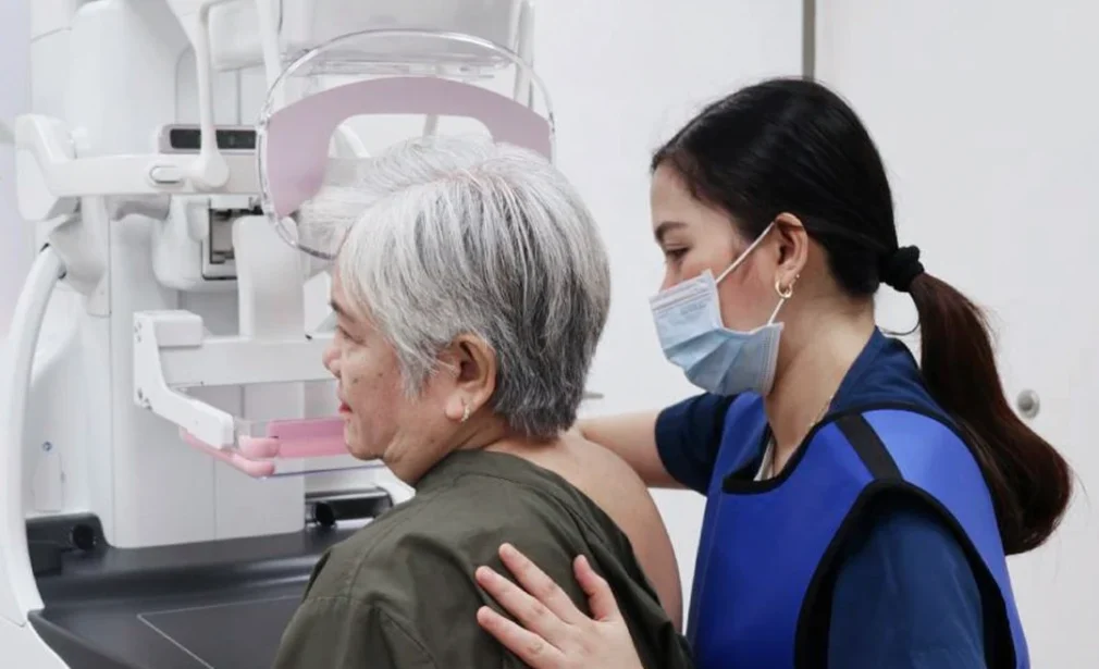
Utilizing cutting-edge technology, this technique captures a series of thin cross-sectional images, enabling a three-dimensional reconstruction of breast tissue. By eliminating the superimposition of structures that can obscure abnormalities, it enhances the detection of subtle lesions, calcifications, and early signs of breast cancer.
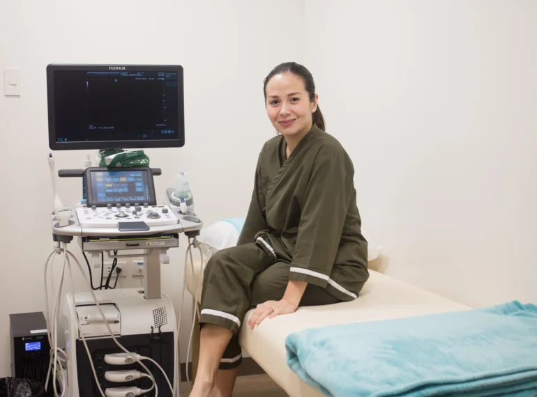
It is highly recommended to undergo both ultrasound and mammography for greater accuracy and detection of breast cancer in women.
Testing Time: 30 Minutes
Recommended for
For women aged 40 and older as part of routine breast cancer screening.
Women under 40 who are at high risk, such as those with a family history of breast cancer or existing breast abnormalities.
Women experiencing breast symptoms, including lumps, pain, nipple discharge, or changes in breast size or shape.
Patients who have had previous breast symptoms or abnormalities may require follow-up screening.
Benefits
Can detect breast cancer before symptoms appear, significantly increasing the chances of successful treatment.
It can identify tumors as small as a few millimeters, which may not be detectable during a physical examination.
Tracks changes in breast tissue over time, making it easier to identify abnormalities at an early stage.
Detect benign conditions such as cysts and calcifications, providing reassurance and guiding further evaluation if necessary.
Book an appointment now

Inquire

Book

Confirm
Best Paired With
Frequently asked questions
You need to secure a doctor’s request before undergoing an Mammography 3D examination. This ensures that the procedure is appropriate for your health condition. If you do not have a request, you may consult with our doctors at Shinagawa. Once the doctor’s request is secured, you can proceed with the procedure.
Mammography 3D can cause discomfort for some individuals, but it is not usually described as painful.
The examination typically lasts for about 20 to 30 minutes, depending on the patient’s condition.
It is recommended to schedule your Mammography 7 to 14 days after the first day of your menstruation.
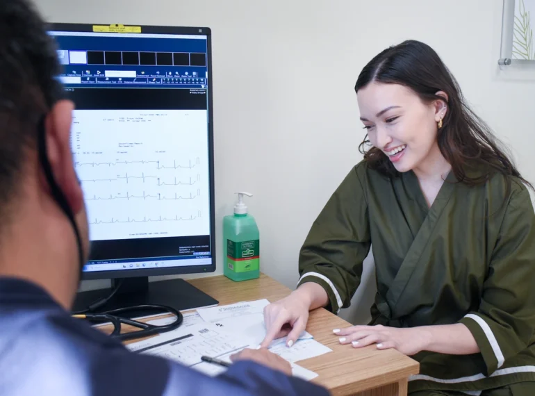
You will receive your results via email within 7 working days, as they will be double-read by our Japanese medical experts. This process ensures the highest level of accuracy in the interpretation of your results. If you need a hard copy, you may request it in advance when booking an appointment through our Patient Care Line. If you have already completed your appointment and need a copy, you may request it here.
Patients aged 40 and above are strongly encouraged to undergo mammography. Women under 40 with a high risk of breast cancer can also do Mammography, including those with a family history of breast cancer, or with breast abnormalities.
Patients should avoid using deodorant, antiperspirant, powders, lotions, creams, or perfumes under the arms, or on or under the breasts the day of appointment.


