

A brain aneurysm is often called a “silent” condition because many people do not realize they have one until it causes serious complications. As a result, it is often perceived as sudden and unpredictable when in reality, many brain aneurysms are associated with identifiable risk factors and can remain undetected for years.
Understanding what causes a brain aneurysm and recognizing its warning signs can help you better understand your risk and the importance of early detection.
What is a Brain Aneurysm?
A brain aneurysm is a weakened area in the wall of a blood vessel in the brain. Over time, blood pressure pushes against this weakened section, causing it to bulge outward like a balloon.
Many cerebral aneurysms remain small and never cause symptoms. Some are only discovered accidentally during imaging performed for unrelated medical concerns. However, if an aneurysm grows larger or ruptures, it becomes a medical emergency that requires immediate treatment.
Not every brain aneurysm will rupture. In fact, many people live their entire lives without knowing they have one. Nevertheless, understanding your personal risk factors and seeking appropriate screening when recommended can help prevent potentially devastating concerns.
What Causes a Brain Aneurysm to Develop?
There is no single answer to what causes brain aneurysm. Instead, most aneurysms develop due to a combination of inherited vulnerabilities and lifestyle-related factors that weaken blood vessel walls over time.
Inherited Risk Factors (Genetics)
Some individuals are naturally more susceptible because of their genetics or underlying medical conditions.
Inherited risk factors and medical contributors include:
- Family history of brain aneurysms, particularly among first-degree relatives
- Certain inherited connective tissue disorders that weaken blood vessel walls
- Congenital abnormalities affecting blood vessel structure
- Genetic conditions associated with vascular abnormalities, such as polycystic kidney disease
- Advancing age, especially after age 40
- Aneurysms occurring more frequently in women
Acquired Risk Factors (Lifestyle Over Time)
Many cases are associated with risk factors that develop gradually throughout life. Common lifestyle habits include:
- High blood pressure (hypertension)
- Smoking
- Excessive alcohol consumption
- Recreational drug use
Recognizing the Symptoms: Unruptured vs. Ruptured Aneurysms
Early Symptoms of a Brain Aneurysm (Unruptured)
Many small aneurysms cause no noticeable symptoms. However, as an aneurysm enlarges, it may begin pressing against nearby nerves or brain structures.
Possible early symptoms of brain aneurysm include:
- Persistent localized headache
- Pain above or behind one eye
- Blurred or double vision
- Dilated pupil
- Drooping eyelid
- Facial numbness or weakness
- Difficulty focusing
- Changes in vision
Ruptured Aneurysm Symptoms
A ruptured aneurysm causes bleeding around the brain, known as a subarachnoid hemorrhage. This is a life-threatening medical emergency.
Symptoms typically appear suddenly and may include:
- An extremely severe headache, often described as the “worst headache of my life”
- Sudden nausea and vomiting
- Stiff neck
- Sensitivity to light
- Blurred vision
- Double vision
- Seizures
- Confusion
- Difficulty speaking
- Weakness or paralysis
- Loss of consciousness
Diagnosing an Aneurysm: Why an MRI or MRA is Essential
Due to the nature of how brain aneurysms develop, imaging plays an essential role in diagnosis. Several imaging tests may be used depending on the patient’s condition, symptoms, and medical history.
Among the most valuable non-invasive tools are:
- Magnetic Resonance Imaging (MRI)
- Magnetic Resonance Angiography (MRA)
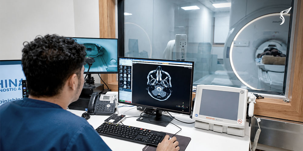
An MRI provides detailed images of the brain’s soft tissues, helping physicians evaluate abnormalities that may be causing neurological symptoms.
An MRA is a specialized MRI technique designed specifically to visualize blood vessels. It allows doctors to examine arteries within the brain and identify possible aneurysms, narrowing, or other vascular abnormalities without requiring invasive procedures in many cases.
Depending on the situation, physicians may also recommend CT scans, CT angiography (CTA), or conventional cerebral angiography to obtain additional information. The choice of imaging depends on the urgency of the situation and the individual’s clinical condition.
Most importantly, imaging should always be interpreted alongside a comprehensive health checkup.
Get Your Health Checkup at Shinagawa Diagnostic
While not every brain aneurysm can be prevented, many of its risk factors can be identified and managed through regular checkups.
Routine health checkups help monitor conditions such as hypertension, diabetes, high cholesterol, and other cardiovascular concerns that may contribute to blood vessel damage over time. For individuals with a family history of a brain aneurysm or other risk factors, advanced imaging may also help identify any abnormalities before any symptoms or severe complications occur.
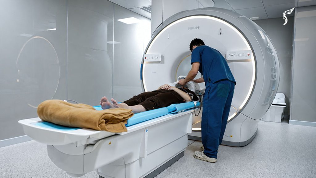
At Shinagawa Diagnostic & Preventive Care, our comprehensive health checkups are designed to provide a thorough assessment of your overall health by providing essential services such as MRI.
In particular, our VIP Shinagawa Dock includes a Brain MRI + Brain MRA + Neck MRA, allowing our medical team to evaluate both brain structures and the blood vessels supplying the brain.
To support accurate and reliable results, we utilize helium-free MRI technology, which delivers high-quality imaging while promoting more sustainable operations. Every MRI examination also undergoes our double-reading method, where findings are independently reviewed by both Filipino radiologists and Japanese medical experts to provide an added layer of quality assurance.
References:
Brain aneurysm. Brain Aneurysm Symptoms, Diagnosis, and Treatment | Brigham and Women’s Hospital. (n.d.). https://www.brighamandwomens.org/neurosurgery/brain-aneurysm
Causes & risk factors. Brain Aneurysm Foundation. (2025, February 12). https://www.bafound.org/understanding-brain-aneurysms/causes-risk-factors/