In an effort to make Japanese-standard health care more accessible to Filipinos, Shinagawa Diagnostic & Preventive Care has partnered with the Civil-Military Operations Regiment (CMOR).
On April 18, this significant event took place at the Ore Central Tower in BGC. Among those who attended the contract signing ceremony were BGEN Benjamin L. Hao, PA, the Regiment Commander of CMOR, and Ms. Masako Uemori, President of Shinagawa Philippines.
This partnership is momentous for both parties as Shinagawa offers both essential and advanced diagnostic services to CMOR members such as the VIP Shinagawa Dock that promotes an in-depth screening focusing on digestive health. This would help in ensuring that the health of Filipino military officers are always in top condition to better serve the country.
Moving forward, may this partnership also be a stepping stone for a better Filipino community that thrives in both health and peace.
https://shinagawa-healthcare.ph/wp-content/uploads/2024/04/CMOR-Cover.webp353940Shinagawa Diagnostic & Preventive Carehttps://shinagawa-healthcare.ph/wp-content/uploads/2024/04/SHSC-BLUE-LOGO-300x70.pngShinagawa Diagnostic & Preventive Care2024-04-26 11:45:202024-04-26 13:45:15
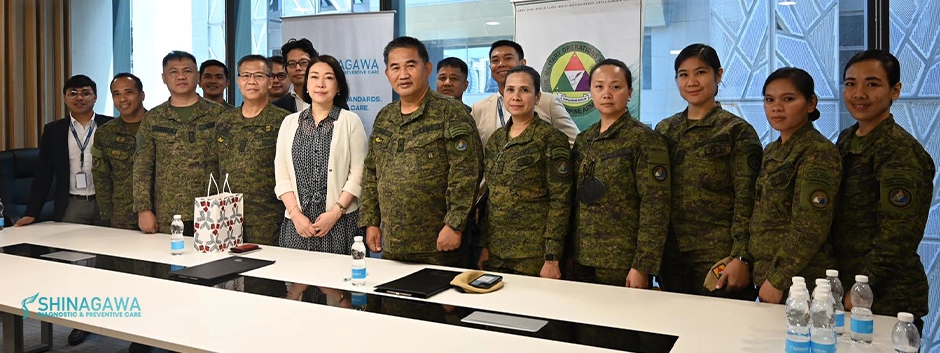
Shinagawa Partners with Civil-Military Operations Regiment (CMOR) for a better Filipino Community
Ever curious about what a 2D echo, or heart ultrasound, can reveal about your heart’s inner workings? This painless procedure goes far beyond a simple picture. It acts as a powerful tool, providing a detailed look at your heart’s health, function, and any potential issues.
In this article, we will explain the world of 2D echo in a way that is simple, informative, and free of technical terminology. We’ll go over why it’s important, how it’s used, and answer any questions you may have about this useful heart health tool.
What is 2D echo?
2D echocardiography, popularly called 2D echo, is a non-invasive test used to analyze the functioning and assess the sections of your heart. This test gives images of the different parts of the heart with the help of sound vibrations. It assists in checking damages, blockages, and blood flow rate. Doppler signals can be colour-coded to enhance visualisation of blood flow (termed Doppler colour-flow mapping) and is the best way to determine the degree of narrowing, calcification or leakage of a valve.
Doctors recommend regular 2D echo tests to analyze and treat any heart issues at the early stages, keeping you healthy and active as you grow old.
Why should you undergo a 2D echo?
An echo test can allow your health care team to look at your heart’s structure and check how well your heart functions. The test helps your health care team find out:
The size and shape of your heart, and the size, thickness and movement of your heart’s walls.
How your heart moves during heartbeats.
The heart’s pumping strength.
If the heart valves are working correctly.
If blood is leaking backwards through your heart valves.
If the heart valves are too narrow.
If a tumor or infectious growth is around your heart valves.
Applications of 2D Echo
2D echo plays a vital role in diagnosing and monitoring a variety of heart conditions. Here’s a breakdown of its key applications:
Diagnosing:
Valve Disorders: can assess the opening and closing of heart valves, helping diagnose issues like stenosis (narrowing) or regurgitation (incomplete closure).
Congenital Heart Defects: It can detect structural abnormalities in the heart chambers, valves, or major blood vessels present at birth.
Heart Failure: helps evaluate the pumping function of the heart chambers and identify signs of heart failure.
Arrhythmias (irregular heartbeats): While not a direct diagnosis tool for arrhythmias, it can identify underlying heart conditions that might contribute to them.
Pericardial Effusion: This refers to fluid buildup around the heart, which it can detect.
Myocarditis: Inflammation of the heart muscle can be visualized through abnormalities in heart wall movement.
Blood Clots: can sometimes help identify blood clots within the heart chambers.
Monitoring:
Tracking Progression of Heart Disease: allows doctors to monitor changes in heart function over time for conditions like heart failure or valve disease.
Pre-surgical Evaluation: Before heart surgery, provides valuable information about the heart’s structure and function to aid surgical planning.
Post-treatment Monitoring: can be used to evaluate the effectiveness of heart medications or interventions like valve replacements.
This list covers some of the most common applications of 2D echo. Remember, a qualified healthcare professional will interpret the results in the context of your individual medical history and symptoms.
Understanding 2D Echo Results
The results of a 2D echo test won’t be a simple image or number. Instead, a trained professional, usually a cardiologist, will analyze the images and measurements obtained during the exam to create a comprehensive report
Interpreting Images
The 2D echo generates images of your heart in real-time, but interpreting these images goes beyond a simple picture. Trained professionals, typically cardiologists with expertise in echocardiography, analyze the images meticulously.
Experience: Cardiologists have extensive training in interpreting echocardiogram images. They can identify subtle abnormalities in size, shape, movement, and blood flow patterns.
Context: They consider these findings in the context of your medical history, symptoms, and other test results to form a complete picture of your heart health.
Integration: The images are just one piece of the puzzle. Cardiologists integrate them with other findings to arrive at an accurate diagnosis.
Measurements and Calculations
A 2D echo goes beyond just images. It also involves obtaining specific measurements and performing calculations to assess your heart’s function with greater precision.
Here’s a breakdown of what the results of a 2D echo test might show:
Measurements
Chamber size: The dimensions of your heart chambers, particularly the ventricles and atria, are measured. Abnormalities in size can indicate various conditions.
Wall thickness: The thickness of your heart muscle walls is measured. Abnormal thickening or thinning can be signs of underlying issues.
Calculations
Ejection Fraction (EF): This is a crucial calculation that reflects the percentage of blood ejected from the left ventricle with each heartbeat. A low EF indicates weakened pumping ability, potentially due to heart failure.
Valve area: Calculations are performed to estimate the size of your heart valve openings. This helps determine if the valves are narrowed (stenosis) or leaking (regurgitation).
Doppler velocities: Doppler echocardiography measures blood flow velocities within the heart. These velocities are used in calculations to assess valve function and blood flow patterns.
How Your 2D Echo Results Pave the Way Forward
The results of your 2D echo act as a roadmap for your heart health journey. A qualified healthcare professional, usually a cardiologist, will analyze the findings and determine the next steps. Here’s how your 2D echo results might guide further action:
Confirmation of Diagnosis: If you have suspected heart problems, a normal 2D echo can be reassuring and may not require further testing. Conversely, abnormal results can confirm a suspected diagnosis like valve disease or heart failure.
Additional Tests: Depending on the findings, your doctor might recommend further tests for a more comprehensive picture. This could include blood tests, stress tests, or cardiac catheterization.
Treatment Plan Development: The results will guide the development of a personalized treatment plan. This might involve medications, lifestyle changes (diet, exercise), or procedures like valve replacement or angioplasty.
Monitoring: Even with normal results, regular follow-up 2D echoes might be recommended, especially if you have risk factors for heart disease. This allows early detection of any potential issues.
Frequently Asked Questions about 2D Echo
Here are some of the most common questions about 2D echo, along with clear and concise answers:
Is a 2D Echo painful?
No, a 2D echo is generally not a painful procedure. You might feel some slight pressure on your chest as the technician presses the transducer to capture images. However, this discomfort is minimal and temporary.
Are there any risks involved?
A 2D echo is a very safe procedure with minimal risks. There’s no radiation involved, unlike some other imaging techniques. In rare cases, some people might experience mild discomfort from lying still for the duration of the exam.
How long does a 2D Echo take?
The actual image capture for a 2D echo typically takes around 20 to 30 minutes. However, the entire appointment, including preparation and explanation of results, might take closer to an hour.
What should I expect after the exam?
There’s no special recovery time needed after a 2D echo. You can resume your normal activities immediately. The technician will remove the gel used during the exam, and you can dress and leave. Your doctor will contact you with the results and discuss any further steps.
Conclusion – 2D Echo for Your Heart
The 2D echo, a painless and non-invasive ultrasound procedure, has emerged as a vital tool in safeguarding your heart health. It offers a window into the inner workings of your heart, revealing valuable insights about its size, shape, function, and potential issues.
By analyzing the images, measurements, and calculations obtained from a 2D echo, doctors can:
Diagnose a wide range of heart conditions.
Monitor the progression of existing heart problems.
Develop personalized treatment plans based on your specific needs.
Remember, a healthy heart is the cornerstone of overall well-being. If you have any concerns about your heart health, a 2D echo can be a valuable first step. Don’t hesitate to discuss your concerns with your doctor. They can assess your individual risk factors and determine if a 2D echo is right for you. Early detection and intervention are key to maintaining a healthy heart for years to come.
Empower Yourself with Knowledge and Action
Empowering yourself with knowledge is vital for maintaining a healthy heart.
Remember, knowledge is power, but action is key. If you have any concerns about your heart health, don’t hesitate to schedule a check-up with your doctor. Early detection and intervention are crucial for preventing and managing heart disease. Take charge of your heart health today!
https://shinagawa-healthcare.ph/wp-content/uploads/2024/04/Importance-Applications-and-Explanations-of-2D-Echo.webp353940Shinagawa Diagnostic & Preventive Carehttps://shinagawa-healthcare.ph/wp-content/uploads/2024/04/SHSC-BLUE-LOGO-300x70.pngShinagawa Diagnostic & Preventive Care2024-04-24 15:39:422024-05-06 08:15:44Importance, Applications and Explanations of 2D Echo
Are you aware of the importance of pneumococcal vaccinations? If not, don’t worry; we’ve got you covered! Here are essential facts to help you understand why getting vaccinated is crucial for your health.
What Are Pneumococcal Vaccinations?
Pneumococcal vaccinations are shots designed to protect individuals from infections caused by Streptococcus pneumoniae bacteria. These bacteria are responsible for various serious illnesses, including pneumonia, meningitis, and bloodstream infections. Booster against hazardous bacteria, such as pneumococcal vaccines, help people bolster their defenses against illness-causing bacteria.
Who Needs Pneumococcal Vaccinations?
Even though everyone benefits from pneumococcal vaccinations, some populations are more susceptible to pneumococcal infections than others. Individuals over 65 who suffer from conditions such as diabetes, heart disease, or weakened immune systems are referred to as having these conditions. Vaccination is crucial for individuals who are at risk of life-threatening illnesses, offering an additional layer of protection.
Two Main Types of Vaccine: Prevenar 13 and Pneumovax 23
There are two primary pneumococcal vaccines available: Prevenar 13 and Pneumovax 23. Both vaccines are designed to prevent pneumococcal diseases, but they work differently and target different strains of Streptococcus pneumoniae.
Prevenar 13
Prevenar 13 is a vaccine that protects individuals from certain infectious diseases by immunizing them. It does this by preparing the body’s defenses to fight the infection, before you catch the bacteria or virus.
Prevenar 13 is a polysaccharide mixture derived from 13 different strains or serotypes of Streptococcus pneumoniae bacteria. Each serotype is joined to a non-toxic protein to make it work more effectively.
Pneumovax 23
is a vaccine recommended for active booster to prevent pneumococcal disease caused by the 23 serotypes contained in the vaccine (1, 2, 3, 4, 5, 6B, 7F, 8, 9N, 9V, 10A, 11A, 12F, 14, 15B, 17F, 18C, 19F, 19A, 20, 22F, 23F, and 33F).
How Do They Work?
Prevenar 13 and Pneumovax 23 function by stimulating the body’s immune response. Vaccines, when administered, introduce harmless bacteria components to the immune system, triggering the production of antibodies against them. This process trains the immune system to recognize and combat bacteria in the future, preventing infection and related illnesses.
Vaccination Schedule
For adults 65 and older, the recommended pneumococcal vaccine schedule usually includes both Prevenar 13 and Pneumovax 23. Prevenar 13 is usually administered first, followed by Pneumovax 23 after a specified interval. Adhering to this schedule ensures maximum effectiveness of the vaccines and provides long-term protection against pneumococcal infections.
What are the Possible Side Effects of Pneumococcal Vaccinations
Typical reported events associated with PCV13 were pain, redness, and swelling at the injection site; limited movement of the injected arm; tiredness; headache; chills; decreased appetite; generalized muscle pain; and joint pain. Similar reactions were observed in adults who received PPSV23.
Effectiveness and Duration of Protection
Both Prevenar 13 and Pneumovax 23 are highly effective at preventing pneumococcal infections. However, the duration of protection offered by these vaccines may vary. Booster doses may be recommended for individuals with underlying health conditions or those at increased risk of bacteria exposure.
Case Study regarding the Safety Data of the Vaccines
In a Case Study conducted (Jackson et al ; Jackson et al) conducted in Europe and the United states with Adults ages ranging from 50 years old and older. Similar reactions have been observed to those adults who have been administered with the vaccine.
Shinagawa Diagnostic & Preventive Care: Your Partner in Health
At Shinagawa Diagnostic & Preventive Care, we prioritize your health and well-being. Our experienced healthcare professionals offer comprehensive vaccination services, including pneumococcal vaccinations, tailored to your individual needs. Schedule a consultation with us today and take proactive steps towards protecting yourself against pneumococcal diseases.
https://shinagawa-healthcare.ph/wp-content/uploads/2024/04/Facts-You-Need-to-Know-About-Pneumococcal-Vaccinations.webp353940Shinagawa Diagnostic & Preventive Carehttps://shinagawa-healthcare.ph/wp-content/uploads/2024/04/SHSC-BLUE-LOGO-300x70.pngShinagawa Diagnostic & Preventive Care2024-04-04 15:28:292024-04-04 15:28:30Facts You Need to Know About Pneumococcal Vaccinations
Medical School
Doctor of Medicine, University of the East, Ramon Magsaysay Memorial Medical Center, Quezon City
Internship
DOH Philippine Centers for Specialized Health Care Integrated Internship Training Program, Quezon City
Residency
Capitol Medical Center, Quezon City
Fellowship
St. Luke’s Medical Center – Quezon City
Affiliations
Philippine College of Cardiology, Diplomate
Philippine College of Physicians, Fellow
Local Sub Specialty Board
Philippine Specialty Board of Adult Cardiology
Philippine Specialty Board of Internal Medicine
JULIUS YVES D. BAGACAY, RMT, M.D., FPCR
Specialty
Radiology
Medical School
San Beda College of Medicine
Internship
Residency
St. Luke’s Medical Center – Quezon City
Fellowship
VRP Medical Center
Certifications
Board Certified Radiologist
Affiliations
Consultant Radiologist, Aventus Medical Care Teleradiology
Consultant Radiologist, Hi-Precision Diagnostics Ortigas
Membership
Member, European Society of Radiology
Local Specialty Board
Philippine Medical Association (PMA)
Local Sub Specialty Board
Philippine Board of Radiology
Philippine College of Radiology
MARIA THERESA S. BUENAFLOR, M.D., FPCR
Specialty
Breast Radiologist
Medical School
University of Santo Tomas
Internship
St. Luke’s Medical Center – Quezon City
Residency
St. Luke’s Medical Center – Quezon City
Fellowship
St. Luke’s Medical Center – Quezon City
Certifications
Contrast-Enhanced Spectral Mammography (2014)
Digital Breast Tomosynthesis (DBT) Interpretation and Performing Tomosynthesis-guided Biopsy (2015 and 2016)
Affiliations
Breast Radiologist, Asian Breast Center, Centuria Medical Makati
Breast Radiologist, Women’s Center, Health Cube Advanced Medical Imaging Unit
Memberships
Corresponding Member, European Society of Radiology (ECR)
International Member, Korean Society of Radiology (KSR)
International Member, European Society of Medical Oncology (ESMO)
Local Specialty Board
Philippine Medical Association (PMA)
Local Sub Specialty Board
Philippine College of Radiology (PCR)
CELINA CELESTE H. ADRANEDA, M.D, FPCP, FPSG
Specialty
Internal Medicine – Gastroenterology
General Hepatology
Medical School
University of Santo Tomas
Internship
University of Santo Tomas
Residency
Cardinal Santos Medical Center
Fellowship
Cardinal Santos Medical Center
Affiliations
Consultant., Cardinal Santos Medical Center
Consultant, Delos Santos Medical Center – Metro Pacific Hospital
Consultant, Jose Reyes Memorial Medical Center
TRIFON ANTONIO D. CABALLERO, M.D., FPSVM, FPCC, FPCP
Specialty
Cardiology
General Medicine
Internal Medicine
Primary Care
Vascular Medicine
Medical School
Pamantasan ng Lungsod ng Maynila
Internship
Ospital ng Maynila Medical Center
Residency
Ospital ng Makati
Fellowship
St. Luke’s Medical Center – Quezon City
Affiliations
Marikina Doctors Hospital and Medical Center
ACE – QC
St. Camillus Medical Center
Arans Medical and Diagnostic Clinic
Memberships
Member, FEU-NRMF Medical Technology Alumni Society
Member, PLM-CM Alumni Society
Member, Rotary Club Manila
Member, Brotherhood of Medical Scholars
Member, Philippine National Red Cross
Member, Ayala Foundation
Local Specialty Board
Philippine Medical Association (PMA)
Local Sub Specialty Board
Philippine College of Cardiology
Philippine College of Physicians
Philippine Society of Vascular Medicine
Philippine Heart Association