
When most people think of eye exams, they imagine reading letters off a chart, checking prescriptions for glasses, or perhaps screening for cataracts or glaucoma. But one of the most important tools in ophthalmology is the fundus examination, also called fundus photography or a retinal exam. This simple, non-invasive procedure allows doctors to look into the back of the eye (the fundus), where key structures such as the retina, optic nerve head, and blood vessels can be found.
Because it is an “eye exam,” many assume fundus photography is only useful when someone already has vision problems. In reality, its greatest value often lies in detecting systemic or ocular diseases.
How a Fundus Examination Works
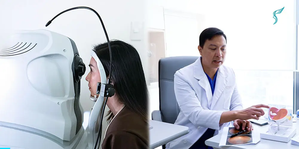
A fundus examination usually begins with the patient seated comfortably, resting their chin and forehead on supports to keep the head steady throughout the procedure.
Before the imaging starts, special eye drops are often applied to dilate the pupils, making them wider and allowing more light to enter the eye. While this is optional, it is commonly done because it helps doctors or technicians capture clearer and more detailed images of the retina and other structures at the back of the eye.
A specialized camera is then used to take high-resolution images of the fundus. During the examination, patients may be asked to look in different directions—up, down, left, or right—so the examiner can assess peripheral areas of the retina.
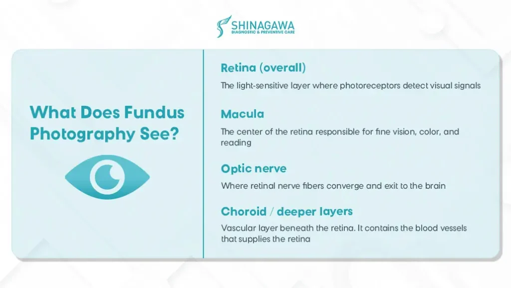
The retina is one of the only internal tissues where blood vessels can be directly seen without surgery. Because of this, the fundus provides a “window” into not just eye health, but vascular and systemic health as well.
What Health Conditions Can a Fundus Exam Detent?
Because the retina is richly vascularized and sensitive to metabolic changes, many of the earliest signs of systemic disease appear in the fundus. Health conditions that may be revealed through a fundus exam include the following:
- Diabetic retinopathy
A complication of diabetes where high blood sugar damages the tiny blood vessels in the retina. Over time, this can cause leakage, bleeding, or scarring, leading to blurred vision or even blindness if untreated.
- Macular edema
A condition where fluid leaks into the macula, the central part of the retina responsible for sharp vision. This swelling blurs central vision and is often linked to diabetic retinopathy or other retinal diseases.
- Hypertensive retinopathy / vascular changes
Caused by high blood pressure, which puts too much strain on the blood vessels in the eyes. This can make them narrow, leak, or thicken, affecting blood flow to the retina and leading to vision problems. It’s also a sign that the blood vessels in the rest of the body may be under stress.
- Glaucoma
A group of eye conditions that damage the optic nerve, often due to increased pressure inside the eye. It develops slowly, without symptoms and can cause permanent vision loss if not detected early.
Get your Fundus Exams at Shinagawa Diagnostic
According to the International Diabetes Federation, the Philippines now has around 4.7 million adults (ages 20–79) living with diabetes.
Since many of the health conditions detected through a fundus exam are closely linked to diabetes, it becomes even more crucial for Filipinos to prioritize early detection and regular screening. With tests like this now more accessible, taking a proactive step can help identify potential complications early—long before symptoms begin to affect vision or overall health.
In countries like Japan, fundus examinations are a common part of routine health checkups, especially for individuals with diabetes risk or those undergoing comprehensive screening programs. In fact, studies have shown that regular checkups which include ocular exams are linked to a lower risk of developing diabetes and hypertension.
By embracing preventive screening, Japan’s healthcare system recognizes this procedure not as an afterthought but as a standard part of routine checkups. Its success in detecting early signs of vascular disease demonstrates what other countries can achieve by adopting the same proactive approach.

Bringing this culture to Shinagawa Diagnostic, we believe that eye health is an integral piece of whole-body wellness. That’s why we include fundus exams as part of our health packages. Just as you get regular blood pressure, cholesterol, kidney, and general imaging tests, adding fundus photography helps complete the picture.
Citations and Resources
Shinagawa Diagnostic & Preventive Care is committed to delivering accurate, reliable, and up-to-date health information. All content published on our platform is grounded in evidence-based research and reviewed by qualified professionals where applicable.
To support our articles, we reference authoritative sources such as peer-reviewed medical journals, official health organizations (e.g., World Health Organization, Department of Health Philippines), and expert guidelines. Where appropriate, we also include data from trusted research institutions and clinical studies.
Our goal is to provide well-researched content that empowers you to make informed choices about your health, diagnostics, and preventive care.
Resources Used in This Article
- Cleveland Clinic. “Fundus Photography, https://my.clevelandclinic.org/health/diagnostics/fundus-photography.”
- National Library of Medicine. “From Bedside to Diagnosis: The Role of Ocular Fundus in Systemic Infections, https://pmc.ncbi.nlm.nih.gov/articles/PMC10707096.”
- Hopkins Medicine. “Diabetes and Your Eyes: What You Need to Know, https://www.hopkinsmedicine.org/health/conditions-and-diseases/diabetes-and-your-eyes-what-you-need-to-know.”