Magnetic Resonance Imaging (MRI) has emerged as one of the most trusted and relied-upon diagnostic tools in modern medicine.
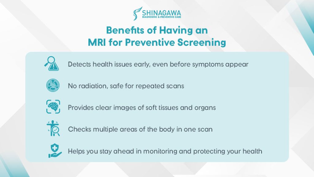
Unlike other imaging exams that depend on ionizing radiation, MRI uses strong magnetic fields and radio waves to generate highly detailed images of the body’s internal structures. This precision makes it especially valuable for early detection and prevention, allowing medical practitioners to identify potential health concerns at their earliest stages.
With its ability to reveal conditions that may not be visible through other imaging methods, it becomes clear why doctors often turn to MRI as their preferred choice in guiding accurate diagnosis and effective treatment.
What is an MRI?
An MRI is a non-invasive imaging test that creates detailed pictures of the inside of the body. Instead of radiation, it uses a powerful magnet and radio waves, which work together to generate clear, cross-sectional images of tissues and organs.
This advanced technology makes MRI especially effective for examining areas that other imaging methods may not capture as precisely, while also offering a safer option for patients who require long-term monitoring or multiple imaging tests over time.
Why would a doctor order an MRI?
A doctor would order an MRI when they need highly detailed images of the body’s soft tissues, organs, and structures that other imaging methods (like X-rays or CT scans) may not show as clearly. Below are some of the most common medical issues that an MRI can help detect:
- Brain and Nerves – to check for tumors, stroke, aneurysms, multiple sclerosis, or nerve damage.
- Spinal Cord and Back – to identify herniated discs, spinal cord compression, or nerve issues.
- Joints and Muscles – to examine torn ligaments, cartilage damage, or sports injuries.
- Heart and Blood Vessels – to detect heart defects, blockages, or vessel inflammation.
- Abdomen and Pelvis – to evaluate the liver, kidneys, uterus, ovaries, prostate, or other internal organs.
What to Expect During an MRI
Preparation for an MRI
- Inform your doctor of implants, pacemakers, or prior surgeries.
- Remove all metallic objects.
- Provide full patient history and symptom evaluation.
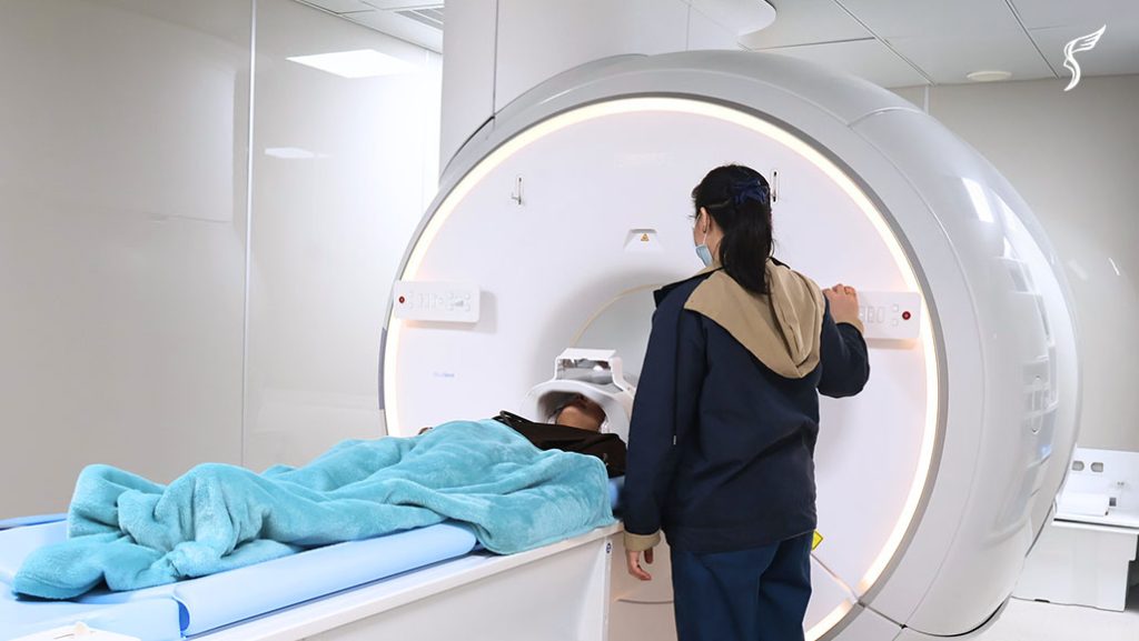
The MRI Machine and Procedure
- The MRI machine is a large, tunnel-shaped device.
- Some patients experience claustrophobia concerns; sedation or open MRI options may help.
- Procedure time: 30–60 minutes.
Safety Concerns
- MR-conditional implants are increasingly safe to scan with, following the ACR Manual on MR Safety 2024 (Source: ACR 2024).
- NHS (2024) confirms MRI can be performed with certain pacemakers under strict monitoring (Source: NHS MRI).
Discover a new standard of MRI care at Shinagawa Diagnostic
When it comes to addressing health concerns that require MRI, having access to the most advanced technology makes early detection far more effective.
At Shinagawa Diagnostic, we have taken this step by introducing the first helium-free MRI technology in the Philippines—delivering clearer images, faster scans, and greater patient comfort, all while being sustainable, reliable, and cost-efficient.
For preventive screening, we focus on non-contrast MRI, which provides detailed images safely without the need for contrast agents, making it ideal for regular health monitoring. Furthermore, to uphold Japanese medical standards, we use a double-reading method in which both Filipino doctors and Japanese medical experts carefully review the results for guaranteed accuracy.
Services under this method include:
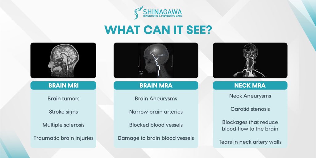
- Brain MRI
- Brain MRA
- Neck MRA
- Brain MRI + MRA
- Brain MRI + Brain MRA + Neck MRA
These are available individually or as part of our comprehensive health package, VIP Shinagawa Dock which combines MRI with a wider spectrum of tests for more thorough check of your body and overall health.
Citations and Resources
Shinagawa Diagnostic & Preventive Care is committed to delivering accurate, reliable, and up-to-date health information. All content published on our platform is grounded in evidence-based research and reviewed by qualified professionals where applicable.
To support our articles, we reference authoritative sources such as peer-reviewed medical journals, official health organizations (e.g., World Health Organization, Department of Health Philippines), and expert guidelines. Where appropriate, we also include data from trusted research institutions and clinical studies.
Our goal is to provide well-researched content that empowers you to make informed choices about your health, diagnostics, and preventive care.
Resources Used in This Article
- Radiology Info. “All About Your Radiology Report: What to Know, https://www.radiologyinfo.org/en/info/all-about-your-radiology-report”
- American College of Radiology. “ACR Appropriateness Criteria, https://www.acr.org/Clinical-Resources/Clinical-Tools-and-Reference/Appropriateness-Criteria”
- Hopkins Medicine. “Magnetic Resonance Imaging (MRI), https://www.hopkinsmedicine.org/health/treatment-tests-and-therapies/magnetic-resonance-imaging-mri#:~:text=MRI%20may%20be%20used%20instead,radiation%20during%20an%20MRI%20procedure.”
- Radiology Info. “Brain MRI, https://www.radiologyinfo.org/en/info/mri-brain”
- National Institute for Health and Care Excellence. “Spinal metastases and metastatic spinal cord compression, https://www.nice.org.uk/guidance/ng234”
- Grand View Research. “U.S. CT And MRI Contrast Media Market Size, Share & Trends Analysis Report By Modality (CT, MRI), By Product (Gadolinium-based Contrast Media, Iodinated Contrast Media), By Application, By Route Of Administration, By End-use, And Segment Forecasts, 2025 – 2030, https://www.grandviewresearch.com/industry-analysis/us-ct-mri-contrast-media-market-report#:~:text=For%20instance%2C%20according%20to%20data,conducted%20annually%20in%20the%20U.S.”
- Jama Network. ‘Projected Lifetime Cancer Risks From Current Computed Tomography Imaging, https://jamanetwork.com/journals/jamainternalmedicine/fullarticle/2832778?guestAccessKey=afde7c2e-df6b-4e7b-9ced-7a15ed74dc1d&utm_source=For_The_Media&utm_medium=referral&utm_campaign=ftm_links&utm_content=tfl&utm_term=041425”
- National Library of Medicine. “Deep learning for the harmonization of structural MRI scans: a survey, https://pmc.ncbi.nlm.nih.gov/articles/PMC11365220/#:~:text=Introduction,1%29.”
- U.S Food and Drug Administration. “FDA Drug Safety Podcast: FDA warns that gadolinium-based contrast agents (GBCAs) are retained in the body; requires new class warnings, https://www.fda.gov/drugs/fda-drug-safety-podcasts/fda-drug-safety-podcast-fda-warns-gadolinium-based-contrast-agents-gbcas-are-retained-body-requires#:~:text=On%20December%2019%2C%202017%2C%20FDA,and%20tissues%20during%20an%20MRI.”
- Radiopaedia. “Diffusion-weighted imaging in acute ischemic stroke, https://radiopaedia.org/articles/diffusion-weighted-imaging-in-acute-ischaemic-stroke#:~:text=Diffusion%2Dweighted%20imaging%20%28DWI%29,the%20benefit%20of%20prior%20imaging.”
- National Institute of Neurological Disorders and Stroke. “Spinal Cord Injury, https://www.ninds.nih.gov/health-information/disorders/spinal-cord-injury#:~:text=Diagnosing%20spinal%20cord%20injuries,fractures%20within%20minutes%20of%20injury.”
- NHS. “Having an MRI scan with an implanted pacemaker or defibrillator, https://www.uhcw.nhs.uk/download/clientfiles/files/Patient%20Information%20Leaflets/Medicine/Cardiology/Having%20an%20MRI%20scan%20with%20an%20implanted%20pacemaker%20or%20defibrillator.pdf”

Inquire

Book